Cell
A cell is the basic, structural and functional unit of life of living organisms. They act as a structural and fundamental unit as many cells together form tissue and many tissues form an organ that forms the organ systems that form the living organisms.
Types of cell:
1. Eukaryotic cells- “Eu” means “true”, while “karyon” means “nucleus” so, a true nucleus is present in eukaryotic cells.
2. Prokaryotic cells- “Pro” means “before”, while “karyon” means “nucleus” so, prokaryotic cells lack a nucleus.
Eukaryotic cell
These are the types of cells present mostly in multicellular organisms such as animals, plants and fungi and some unicellular organisms such as protists.
These cells have specialised membrane-bound structures known as cell organelles and a nucleus. A cell membrane bounds the cells. The cells in some organisms, such as plants, have one more covering outside cell membrane is known as the cell wall.
Let us see the structure of a eukaryotic cell:
i. Cell membrane/ Plasma membrane-
This is a selectively permeable membrane that allows the movement of specific substances in and out of the cell. It is formed of a phospholipid bilayer in which the proteins are embedded.
The fluid mosaic model explains the structure of the plasma membrane. This is because the phospholipid bilayer has a fluid-like consistency, and the structures like proteins and cholesterol are flowing in it like a mosaic. The phospholipid molecules in the bilayer have a hydrophilic (water-loving) head and two hydrophobic tails (water-fearing).
It allows the movement of useful substances such as water, oxygen, and organic molecules into the cells. It removes waste materials like carbon dioxide and nitrogenous waste (ammonia) out of the cell. The cell membrane has receptors on it that help to detect the chemicals like hormones.
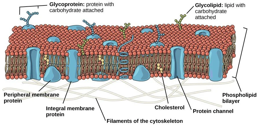
Fig. 1: Structure of plasma membrane
Image source: Openstax
Transport across the cell membrane- The molecules can move in and out of the cell by various methods such as:
1. Diffusion- A method of transport in which molecules other than water moves from the region of higher concentration to the region of low concentration. Oxygen and carbon dioxide can move via this method. This does not need energy (ATP), so it is passive transport.
2. Osmosis- A method of transport in which water moves from the region of higher concentration to the region of low concentration. This does not need energy (ATP), so it is passive transport.
3. Active transport- A type of transport in which the molecules move from the region of low concentration to high concentration and thus needs energy as transport is against the gradient.
ii. Cytoplasm-
The complete area of the cell between the plasma membrane and the nucleus is known as cytoplasm. It has a gel-like cytosol in which the organelles are present.
iii. Nucleus-
It is the largest cell organelle that controls the activity of other organelles of the cell. Its membrane is known as nuclear-envelope and is double-layered. The nucleus contains the chromosomes (chromatin) in which the DNA is present, which controls the activity of the cell.
The nuclear envelope has pores that allow movement of molecules between the nucleus and the cytoplasm. The molecules that move out of the nucleus are RNA and proteins, while the substances that can move inside the nucleus are ribosomes and ATP.
The part of the nucleus in which the RNAs and proteins are aggregated is known as nucleolus. It synthesises ribosomes.
A semi-solid fluid, like cytoplasm, is present inside the nucleus known as nucleoplasm.
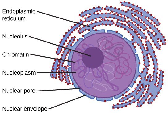
Fig. 2: Structure of eukaryotic Nucleus
Image source: Openstax
iv. Ribosomes-
These are the smallest organelles of the cell that help in the process of protein synthesis in the cell. They are present inside the nucleus, scattered in the cytoplasm and on the membrane of another organelle known as Endoplasmic reticulum. It is made up of RNA and protein. The eukaryotes have 80 S types of the ribosome. It is formed of two subunits: 40 S and 60 S. (prokaryotes have 70 S ribosomes). It is known as the “protein factory” of the cell.

Fig. 3: Structure of Ribosome (Undergoing protein synthesis
Image source: Openstax
iv. Endoplasmic reticulum-
It is a structure connected to the nuclear envelope. It is formed of a network of membranes covering a space filled with fluid.
It is of two types:
a. Smooth endoplasmic reticulum (SER)- The smooth endoplasmic reticulum appears smooth as it does not have ribosomes attached to the membrane. It is involved in the functions of the cell, such as synthesis of lipids and steroid hormones such as estrogen and testosterone. A special SER is present in the muscle cells, which is known as the sarcoplasmic reticulum.
b. Rough endoplasmic reticulum (RER)- The rough endoplasmic reticulum appears rough as it has ribosomes attached to the membrane. It is involved in the modification of the proteins that are synthesised by the ribosomes. The proteins move out of the RER in the vesicles that shed off from the RER.
v. Golgi body/ Golgi apparatus-
It is present adjacent to the endoplasmic reticulum. It looks like a pile of flat sacs presents one over another. These flat sacs are known as cristae.
The vesicles move from the ER to the Golgi apparatus at one end. This end of the Golgi body is known as “cis face.” These contain the lipids and proteins synthesised in the ER. The molecules synthesised in the ER are processed, sorted, and packaged in the Golgi body and are released from another end of the Golgi body as Golgi vesicles. This end of the Golgi body is known as “trans face.” These molecules are tagged by the Golgi body to direct them towards the desired part of the cell.
The Golgi body and Golgi vesicles are together known as the Golgi apparatus or Golgi complex. The Golgi apparatus also make a cell organelle known as lysosomes. In plant cells, they form polysaccharides which are present in the cell wall.
Remember: Nuclear envelope, Endoplasmic reticulum, Golgi apparatus, and plasma membrane are together known as the Endomembrane system.

Fig. 4: Structure of the Endomembrane System
Image source: Openstax
vi. Lysosomes-
These are small and spherical organelles with a single membrane. The lysosomes contain hydrolytic or digestive enzymes. These hydrolytic enzymes help to destroy or breakdown of the unwanted, worn-out components of the cell. Sometimes, they can also digest the complete cell when it is not required. The lysosomes are also present in the white blood cells (macrophages), where they help to destroy the foreign particles or pathogens and help in the immune defence.
A special type of lysosome is present in the head of the sperm, known as acrosome. It helps to break the membrane of the ovum to enter it during fertilisation. These are not present in plant cells.
vii. Mitochondria-
It is an oval-shaped cell organelle with a double membrane. The outer membrane of the mitochondria is smooth, while the inner membrane has folds inwards that are known as cristae. The mitochondria have their own DNA and ribosomes.
It is known as the “powerhouse or energy factories of the cell” as it synthesises energy in the form of ATP molecules.
The part of the mitochondria that is present inside the inner membrane is known as the matrix. The space between the membrane of the mitochondria is known as intermembrane space. The outer mitochondrial membrane is more permeable as it has proteins known as porins that allow entry of small, water-soluble molecules into the intermembrane space. The inner membrane is selectively permeable that allows entry of selected material inside.
ATP is produced by cellular respiration in the cell. In mitochondria, aerobic type of respiration takes place that involves using oxygen. The energy is produced from molecules such as sugars (glucose) and fats. The steps of aerobic respiration take place in the matrix and the inner membrane. The folds of the inner membrane increase the surface area for ATP production. The ATP produced in this process can move out of the mitochondria to reach different parts of the cell.

Fig. 5: (a) Structure of mitochondria (b) Microscopic view of mitochondria
Image source: Openstax
Remember: ATP is the universal energy carrier molecule of the cell.
viii. Peroxisomes-
These are small organelles that perform the beta-oxidation process in the breakdown of fatty acids and amino acids. These also help in the detoxification of toxins and alcohols. Specialised peroxisomes present in the plant cells are known as glyoxysomes. These synthesise glucose by gluconeogenesis from molecules like fats.
ix. Vacuoles-
These are small membrane-bound organelles that store the molecules such as food, nutrients, and waste molecules for a short duration of time. A large central vacuole is present in the plant cell, while numerous small vacuoles are present in animal cells.
x. Centrosome-
This organelle is present near the nucleus of the animal cells. It is the microtubule-organizing centre. It contains two centrioles, present perpendicular to each other. Each centriole is formed of 9 triplets of microtubules. They are involved in cell division and help in the movement of the chromosomes during cell division. These are not present in plant cells.

Fig. 6: Structure of Centrosome
Image source: Openstax
xi. Cell wall-
It is a rigid structure present outside the cell membrane in plant cells. It helps the plant cell to have structural support and also provides the shape to the plant cell. It is also present in some fungi and protist cells but is absent in animal cells. In plant cells, it is formed of cellulose and lignin while in fungal cells, it is formed of chitin.
Remember: Unlike the plasma membrane, the cell wall is a semi-permeable membrane.
xii. Chloroplast-
It is an oval-shaped organelle with a double membrane. It also has its own DNA and ribosomes. It is involved in the photosynthesis of food in the plant cells. It is absent in animal cells. These are known as “food factories of the cells”.
It has an inner membrane, an outer membrane, and intermembrane space. Inside the inner membrane, the stroma is present in which stacks of thylakoids are present. Each stack of the thylakoid is known as grana.
Chlorophyll pigment that helps to absorb light for photosynthesis is present in the chloroplasts. Some protists also have chloroplasts and can synthesise their own food.

Fig. 7: Structure of Chloroplast
Image source: Openstax
Remember: The plant cells appear green due to the presence of chlorophyll pigment.
xiii. Cytoskeleton-
These are the protein fibres that help to maintain the shape of the cell. The types of protein fibres are:
a. Microfilaments: Narrow protein fibres formed of actin globular protein and help in the movement of the cell.
b. Intermediate filaments: These are larger than microfilaments and smaller than microtubules. They help in maintaining the cell shape and keep the cell organelles in their places.
c. Microtubules: These are tube-like structures formed of alpha and beta-tubulin proteins. They help in chromosome movement in the cell during cell division. They also form cilia and flagella, which are hair-like structures present on the plasma membrane and help in the movement of the cell. Cilia is also present in the lining of the respiratory tract and fallopian tube.
Compare plant cell with animal cell:
1. Plant cell contains a cell wall that is absent in the animal cell.
2. Plant cell contains chloroplasts which are absent in animal cells.
3. Plant cell has a single and large central vacuole, while the animal cell has small vacuoles.
4. Plant cell does not have lysosomes that are present in an animal cell.
5. Plant cell does not have centrosomes that are present in an animal cell.
6. Plant cells do not have cilia and flagella that are present in animal cells.

Fig. 8: Structure of a Plant cell
Image source: Openstax

Fig. 9: Structure of an Animal cell
Image source: Openstax